Приложение WiSoft Athena Viral Spread позволяет исследователям, проводящим скрининг тестируемых соединений или антител на предмет вмешательства в распространение вирусов между соседними клетками в монослое, получить новое представление.
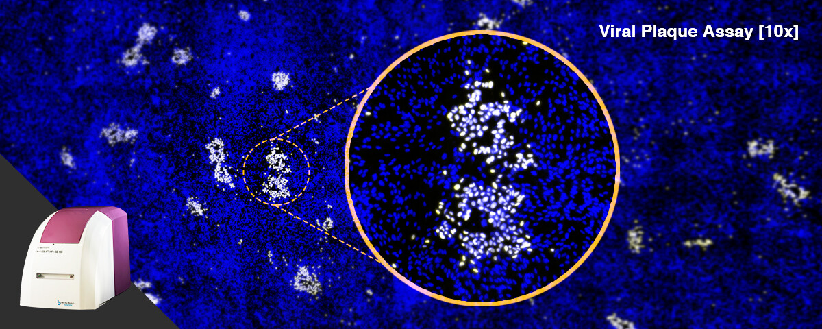
Анализ совместим со сшитыми монтажными изображениями, визуализирующими большие области многолуночных планшетов или посуды, обычно полученными при малом увеличении, с многочисленными метриками.
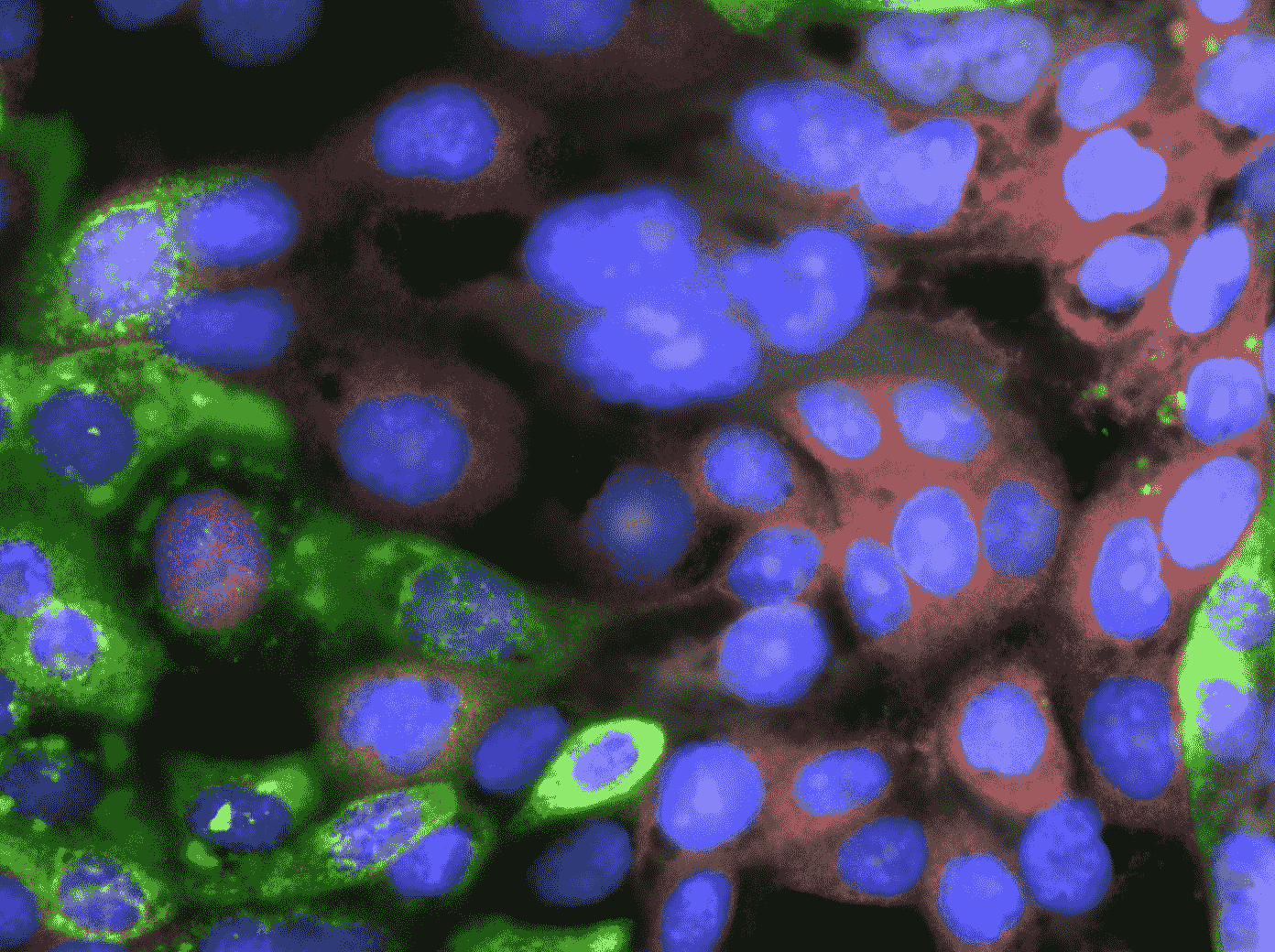
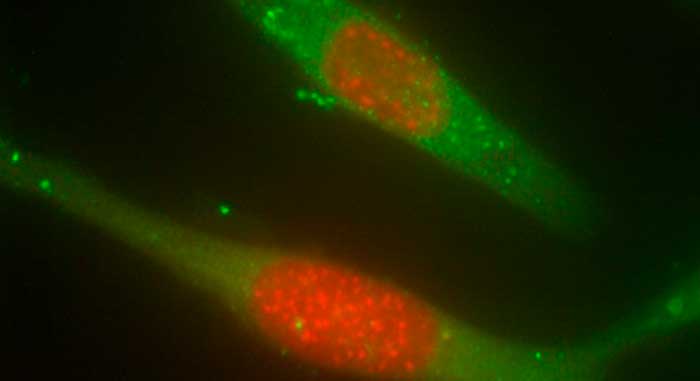
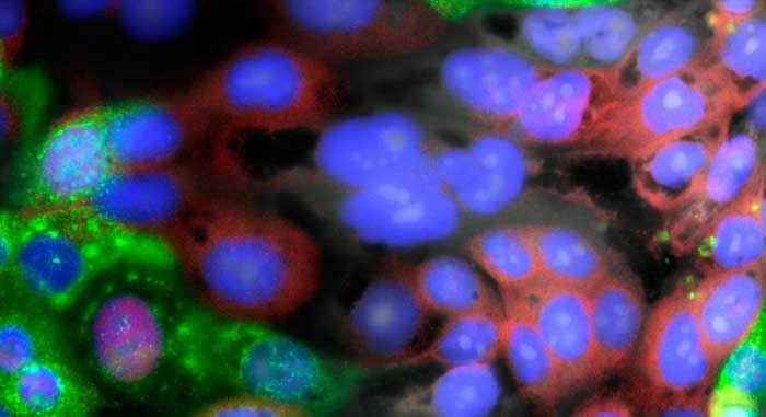
Программное обеспечение сначала идентифицирует клетки по их ядру, а затем определяет их цитоплазматические границы. Внутри каждой клетки используется специфический для инфекции канал, чтобы идентифицировать каждую клетку как инфицированную или здоровую. При желании можно также измерить сигнал флуоресценции в дополнительном канале белка, представляющего интерес (POI), для мониторинга клеточной реакции на инфекцию.
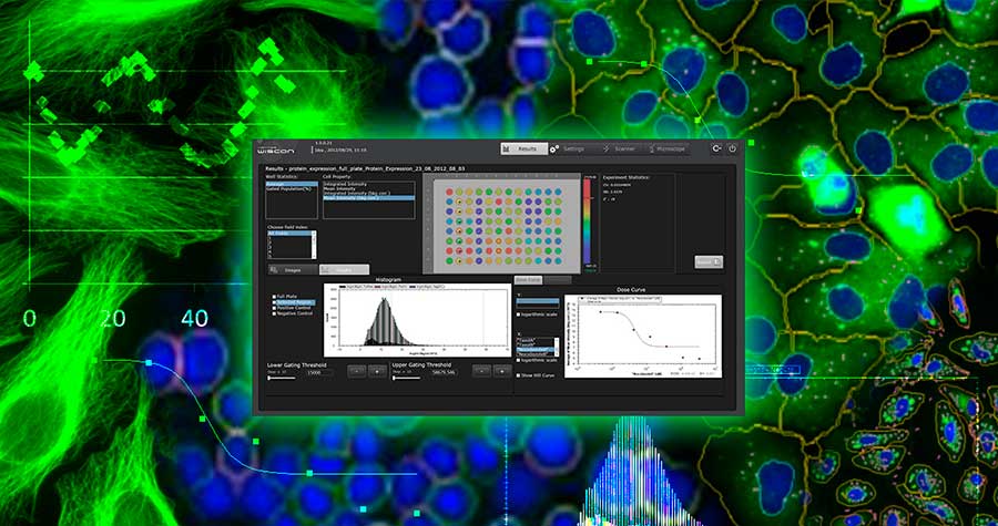
Для каждой клетки приложение измеряет:
Таким образом, исследователи могут изучать изменения в росте и гибели клеток, а также распространение вирусной инфекции с течением времени.
Для каждой клетки приложение измеряет: