Вам никогда не казалось, что вы ищете иголку в стоге сена?
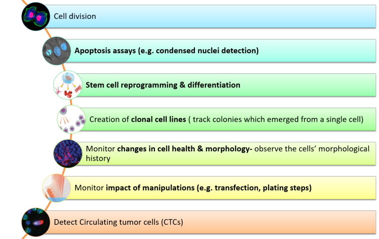
Визуализация редких событий требует систематического, непрерывного отслеживания многомерных аспектов системы, фиксации динамических процессов и выявления переходных, иногда неожиданных особенностей.
Такое отслеживание должно включать автоматическое и высокоточное обнаружение конкретных событий, представляющих интерес, а также возможность быстрого захвата и документирования этих событий с высоким разрешением.
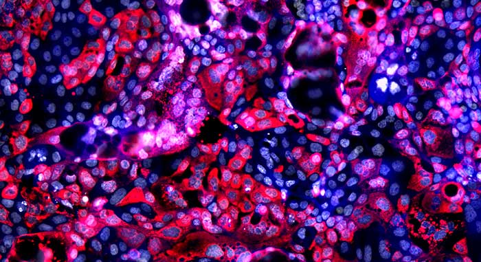
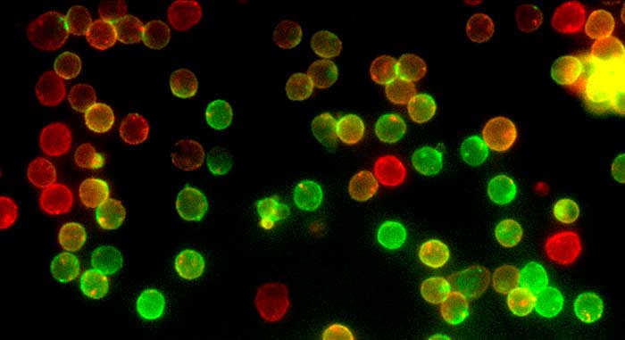
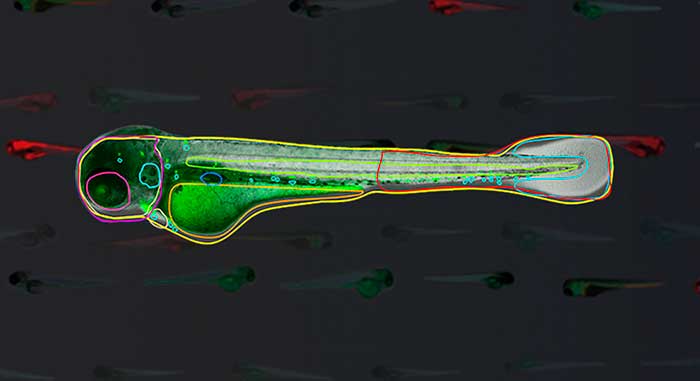
WiScan® Hermes включает в себя сложные инструменты HCS для изучения редких событий в живых клетках.
Просматривайте изображения только тогда, когда происходит что-то интересное, и проверяйте библиотеку изображений, чтобы узнать, что именно произошло.
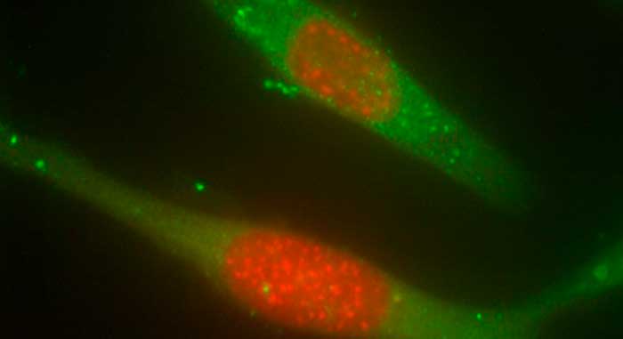
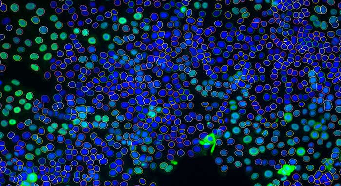
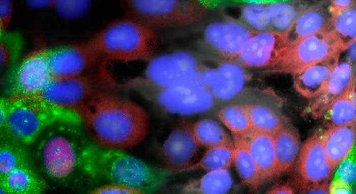
Эта уникальная функция используется для пространственного обнаружения и характеристики временной кинетики редких событий. Используя передовую систему HCS (High Content Screening) компании IDEA Bio-Medical, основанную на сверхбыстрых свойствах микроскопа, система сначала сканирует всю пластину и получает изображения с малым увеличением (2X-10X) через заданные временные интервалы. Это начальное сканирование автоматически выявляет интересующие объекты, основываясь на многопараметрических морфологических признаках или свойствах флуоресценции.
Эта уникальная функция используется для пространственного обнаружения и характеристики временной кинетики редких событий. Используя передовую систему HCS (High Content Screening) компании IDEA Bio-Medical, основанную на свойствах сверхбыстрого получения изображений микроскопа, система сначала сканирует всю пластину и получает изображения с малым увеличением (2X-10X) через заданные временные интервалы. Это начальное сканирование автоматически выявляет интересующие объекты, основываясь на многопараметрических морфологических признаках или свойствах флуоресценции.
Такая сверхбыстрая скорость сканирования достигается благодаря уникальным возможностям системы визуализации Hermes,
которые включают в себя неподвижную пластину (подвижным компонентом является объектив) и многоканальную одновременную визуализацию. Быстрая автоматическая смена
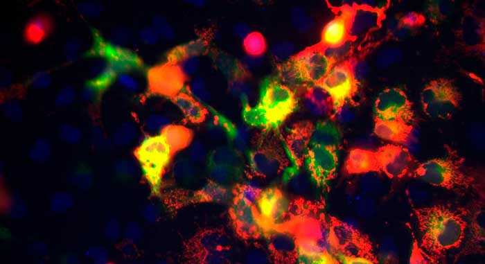
Между сканированиями происходит быстрая автоматическая смена объективов, а затем второе сканирование, при котором получают изображения и записывают видеоролики в больших увеличениях (20X-60X) только для выбранных объектов, представляющих интерес (D), которые точно наводятся и фиксируются благодаря быстрой фокусировке и высокой повторяемости позиционирования сканера микроскопа.