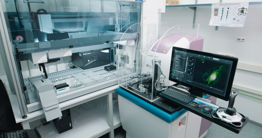
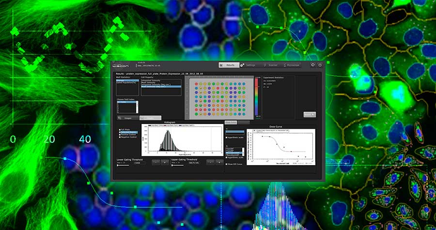
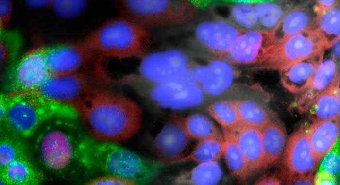
Сопряжение анализа репортерных генов на основе клеток с платформой высококонтентного скрининга Hermes обеспечивает комплексное решение для успешного скрининга транслокации белков из одного клеточного компартмента в другой.
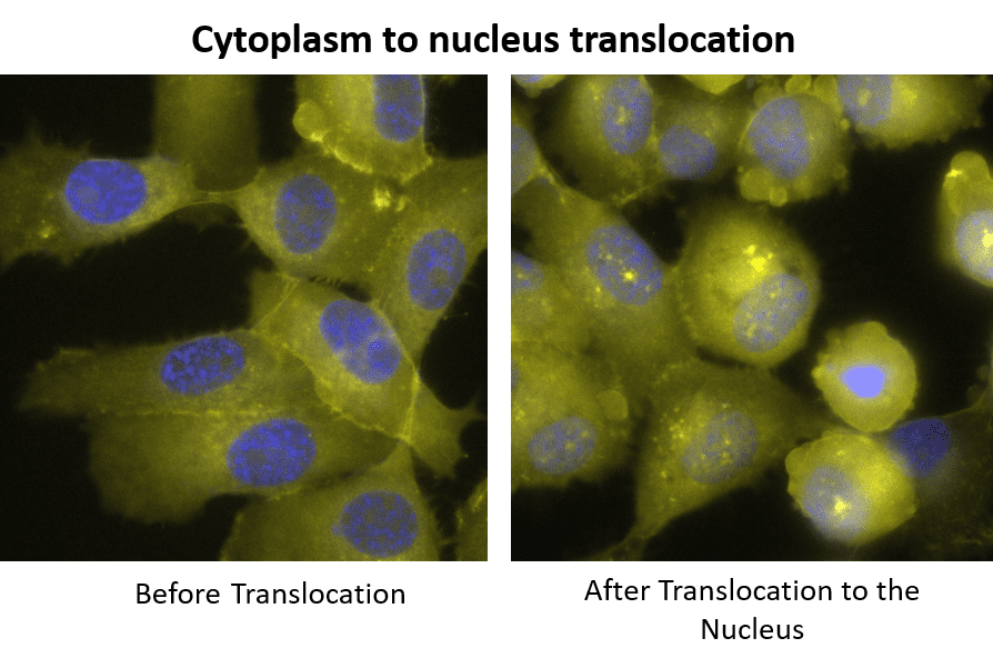
Транслокация белков между клеточными компартментами — ключевой процесс в клеточной биологии. В транслокации участвует множество процессов, включая передачу сигналов для изменения функции клетки (например, экспрессии генов), иммунную стимуляцию при вирусных инфекциях и перенос груза в ядро и из него.
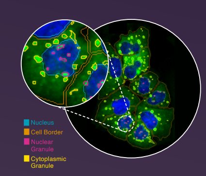
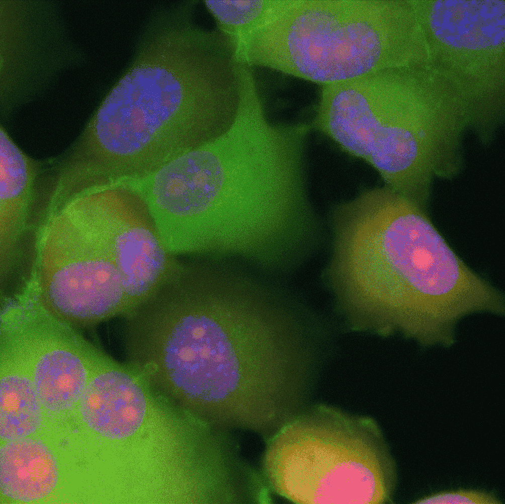
Визуализация является ценным подходом для мониторинга процесса транслокации путем визуализации белка в различных компартментах, измерения его интенсивности и расчета отношения интенсивности меченого белка между цитоплазматическим и ядерным компартментами в качестве количественной меры транслокации.
Данный анализ позволяет измерить соотношение интенсивности меченого белка между цитоплазматическим и ядерным компартментами в качестве количественной меры транслокации.
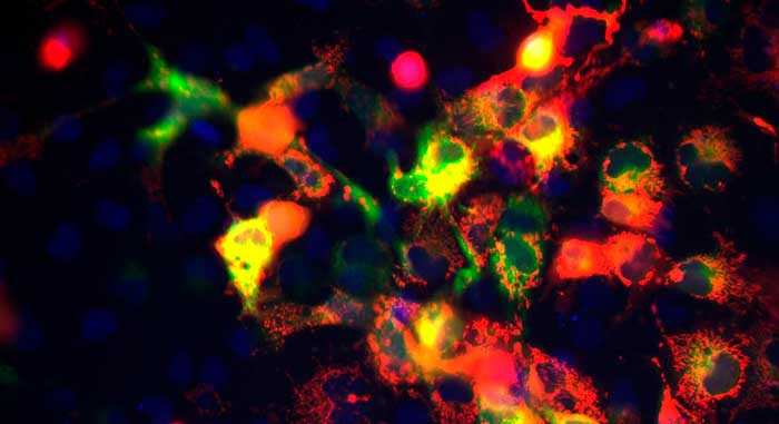
Сопряжение анализа репортерных генов на основе клеток с платформой высококонтентного скрининга Hermes обеспечивает комплексное решение для успешного скрининга транслокации белков из одного клеточного компартмента в другой.
Транслокация белков между клеточными компартментами — ключевой процесс в клеточной биологии. В транслокации участвует множество процессов, включая передачу сигналов для изменения функции клетки (например, экспрессии генов), иммунную стимуляцию при вирусных инфекциях и перенос груза в ядро и из него.
Визуализация является ценным подходом для мониторинга процесса транслокации путем визуализации белка в различных компартментах, измерения его интенсивности и расчета отношения интенсивности меченого белка между цитоплазматическим и ядерным компартментами в качестве количественной меры транслокации.
Данный анализ позволяет измерить соотношение интенсивности меченого белка между цитоплазматическим и ядерным компартментами в качестве количественной меры транслокации.
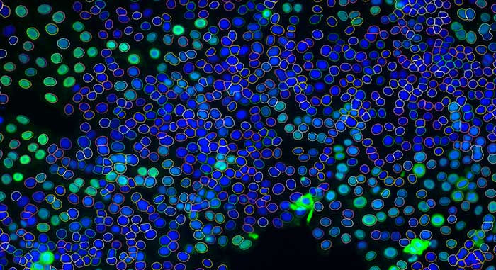
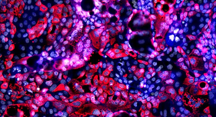
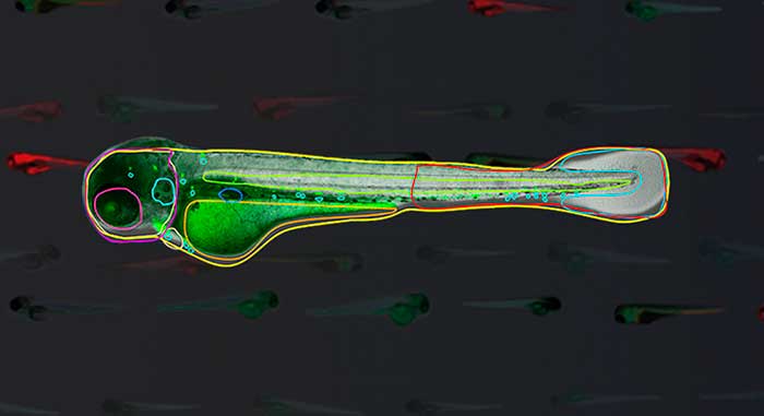
Анализ транслокации ядер с использованием мечения DAPI + FAR RED и 10-кратного увеличения на системе визуализации клеток Hermes. Автоматизированный анализ изображений, выполняемый одновременно с получением, выявляет лунки, демонстрирующие высокую ядерную транслокацию при обработке (красный цвет), по сравнению с низкой транслокацией, измеренной в контрольных лунках (синий цвет).
Tab Content